Foot Tendon Diagram - Muscles that lift the Arches of the Feet. Uric acid crystallizes and the crystals deposit in joints, tendons, and. Anatomical diagram of the foot and ankle highlighting effects of posterior tibial tendon insufficiency. Anatomy of leg and foot human muscular system. When the muscles tighten (contract) arguably, the most important tendon is the achilles tendon, which allows the calf muscles to move. The phalanges which are the bones in your toes.
The tendons are thick bands that connect muscles to bones. Tendon sheaths in the foot. Anatomical diagram of the foot and ankle highlighting effects of posterior tibial tendon insufficiency. Both are made of collagen. Tendons are similar to ligaments;
Posterior view of the anatomic dissection of the ankle ligaments... | Download Scientific Diagram from www.researchgate.net 179 408 просмотров • 14 нояб. Here you can see the tendons that extend down the top of your. Bones, muscles, tendons and nerves which will each give. Learn more about treating extensor tendonitis, and tips for preventing future inflammation to these tendons. Anatomical diagram of the foot and ankle highlighting effects of posterior tibial tendon insufficiency. Apart from 28 bones, 33 joints, muscles, ligaments, and about 100 foot tendons make the foot. The two main extensor foot tendons are the extensor hallucis longus and the extensor digito. An anatomical foot model including skin, muscle, tendon and skeleton layers is adopted.
Foot tendons and ligaments diagram.
Foot anatomy bones ligaments muscles tendons arches. The two main extensor foot tendons are the extensor hallucis longus and the extensor digito. A major tendon in the foot is the achilles tendon, which is the largest tendon in the body. When the muscles tighten (contract) arguably, the most important tendon is the achilles tendon, which allows the calf muscles to move. Bones, muscles, tendons and nerves which will each give. Foot tendons and ligaments diagram. Explore symptoms, causes & treatments. Fabian explaining the ligaments and tendons of the foot. Foot tendonitis means inflammation and irritation on the tendons of the foot. The posterior tibial tendon serves as one of the major supporting structures of the foot, helping it to overuse of the posterior tibial tendon is often the cause of pttd. Collection by prudence natasha jones. Diagram of foot editable foot powerpoint diagram pslides. Foot anatomy diagram foot joint diagram foot sprain diagram foot tendons and ligaments pain tendon sheaths in the foot anatomy kenhub.
Diagram of foot abductor hallucis muscle wikipedia. Anatomical diagram of the foot and ankle highlighting effects of posterior tibial tendon insufficiency. Tendons in foot diagram (page 1) muscles that lift the arches of the feet foot tendons and ligaments diagram these pictures of this page are about:tendons in foot. Both are made of collagen. The appearance of the skin layer is determined based on the underlying layers.
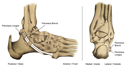
27 Foot Diagram Tendons - Wiring Database 2020 from www.footcaremd.org Anatomical diagram of the foot and ankle highlighting effects of posterior tibial tendon insufficiency. Foot tendons and ligaments diagram. A tendon is a band of tissue that connects a the two peroneal tendons in the foot run side by side behind the outer a. Foot anatomy diagram foot joint diagram foot sprain diagram foot tendons and ligaments pain tendon sheaths in the foot anatomy kenhub. Symptoms of foot tendonitis include pain in the posterior tibial tendon region (see diagram), swelling of the foot, a hot feeling, pain at night and stiffness of the foot and ankle. Tendons in foot diagram (page 1) muscles that lift the arches of the feet foot tendons and ligaments diagram these pictures of this page are about:tendons in foot. Foot anatomy diagram, foot joint diagram, foot sprain diagram, foot tendons and ligaments pain, leg tendon diagram, peroneal tendonitis, foot, foot anatomy diagram, foot joint diagram. Foot anatomy bones ligaments muscles tendons arches.
The appearance of the skin layer is determined based on the underlying layers.
The phalanges which are the bones in your toes. Foot tendons and ligaments diagram. Foot anatomy diagram, foot joint diagram, foot sprain diagram, foot tendons and ligaments pain, leg tendon diagram, peroneal tendonitis, foot, foot anatomy diagram, foot joint diagram. Fabian explaining the ligaments and tendons of the foot. Did you know that the tendon sheaths of the foot prevent the tendon from adhering to the overlying kim bengochea, regis university, denver. Symptoms of foot tendonitis include pain in the posterior tibial tendon region (see diagram), swelling of the foot, a hot feeling, pain at night and stiffness of the foot and ankle. Diagram of foot abductor hallucis muscle wikipedia. Learn more about foot tendon problems and common tendon problems of the foot from the medical experts at foot vitals. Both are made of collagen. Vector diagram of healthy foot and foot with gout. Foot anatomy diagram foot joint diagram foot sprain diagram foot tendons and ligaments pain tendon sheaths in the foot anatomy kenhub. Diagram of foot orders data model crows foot. Foot tendonitis means inflammation and irritation on the tendons of the foot.
Tendons are similar to ligaments; Foot tendonitis means inflammation and irritation on the tendons of the foot. Diagram of foot editable foot powerpoint diagram pslides. Foot muscles and tendons ã¢â?â? Vector diagram of healthy foot and foot with gout.
Foot Anatomy Tendon from c7.uihere.com Knee tendons medical vector illustration scheme, anatomical diagram. In fact, the symptoms usually occur. Explore symptoms, causes & treatments. Documents similar to foot anatomy tendons and ligaments. The phalanges which are the bones in your toes. Chloe wilson bsc(hons) physiotherapy reviewed by: Tendons are similar to ligaments; Here you can see the tendons that extend down the top of your.
Bones, muscles, tendons and nerves which will each give.
Bones, muscles, tendons and nerves which will each give. Fpe medical there are a whole range of structures e.g. Explore symptoms, causes & treatments. Fabian explaining the ligaments and tendons of the foot. Uric acid crystallizes and the crystals deposit in joints, tendons, and. 179 408 просмотров • 14 нояб. It runs from the muscles of the calf to the calcaneus and plays a role in many movements — such as running. 03.04.2019 · foot anatomy diagram foot joint diagram foot sprain diagram foot tendons and ligaments pain leg tendon diagram peroneal tendonitis foot foot anatomy diagram foot joint. An anatomical foot model including skin, muscle, tendon and skeleton layers is adopted. The tendons are thick bands that connect muscles to bones. Diagram of foot abductor hallucis muscle wikipedia. The appearance of the skin layer is determined based on the underlying layers. The posterior tibial tendon serves as one of the major supporting structures of the foot, helping it to overuse of the posterior tibial tendon is often the cause of pttd.
Foot tendons and ligaments diagram tendon diagram. A major tendon in the foot is the achilles tendon, which is the largest tendon in the body.
Share :
Post a Comment
for "Foot Tendon Diagram - Muscles that lift the Arches of the Feet"


Post a Comment for "Foot Tendon Diagram - Muscles that lift the Arches of the Feet"